What Is the New Discovery About Axon Structure?
Question: What did Johns Hopkins researchers discover about axon structure?
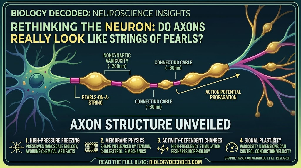
They found that unmyelinated mouse axons can naturally contain repeated nanoscale swellings that make them resemble strings of pearls, rather than simple cylindrical tubes. The researchers called these structures nonsynaptic varicosities, because the swellings were not synapses.
This is important because axons are often introduced as long, cable-like extensions of neurons. That description is useful, but it can make their physical architecture sound much simpler than it really is.
The 2024 Nature Neuroscience study found a thin axonal cable of roughly 60 nanometers between repeated swellings that were about 200 nanometers in diameter. Those dimensions are extraordinarily small,far below the width of a human hair.
Definition + Expansion: What Is an Axon?
An axon is the long projection of a neuron that carries electrical signals away from the cell body toward other cells.
Axons can extend across substantial distances within nervous tissue and are essential for communication between neurons and other cells. Some are covered by myelin, an insulating layer that changes how electrical signals propagate, while others are unmyelinated.
The Johns Hopkins study focused on unmyelinated axons, where the membrane itself is exposed to physical forces that can influence its shape.
The new evidence suggests that understanding an axon’s function may require looking beyond the simplified “wire” analogy.
Why Were Axons Traditionally Drawn as Smooth Tubes?
The traditional illustration is not necessarily wrong as a simplified teaching model.
Scientists have long understood that axon diameter can vary and that axons contain structures such as synaptic varicosities. But the newly described nanoscale pearling is different from those familiar structures.
The problem partly comes down to how biological tissue is prepared for microscopy.
Traditional electron microscopy often uses chemical fixation and dehydration. Those processes can distort delicate membrane structures, potentially making an axon appear more cylindrical than it is under near-native conditions.
Question: Does this mean scientists have been completely wrong about axons for 100 years?
Not exactly. The cylindrical axon remains a useful abstraction, and axons genuinely can appear tubular under many imaging and preparation conditions. The new research instead suggests that this familiar representation may leave out important nanoscale structure, particularly in unmyelinated axons.
Johns Hopkins described the finding as challenging a century of understanding about axon structure, while the researchers’ actual work is more specifically focused on revealing nanoscale morphology that conventional preparation methods could obscure.
That distinction is important.
Science often replaces overly simple models with more detailed ones rather than declaring the earlier model completely useless.
What Does a “Pearls-on-a-String” Axon Look Like?
Picture a very thin biological cable.
Now imagine that instead of having exactly the same diameter throughout its length, the cable repeatedly expands and narrows:
thin section → swollen section → thin section → swollen section → thin section
At microscopic scale, this creates the appearance of beads threaded along a string.
The researchers found that the swellings were approximately 200 nanometers across, while the narrow connector regions were around 60 nanometers in diameter.
The structures were also distinguished from the larger beading associated with damaged or degenerating axons.
Normal Pearling vs Disease-Associated Beading
Beading itself is not new to neuroscience.
Scientists have observed larger axonal swellings in neurons undergoing damage, including in neurodegenerative conditions. Such changes can accompany disruption of the axon’s membrane and internal structural framework.
The Johns Hopkins finding is different because the researchers observed small, repeating swellings in axons under conditions intended to preserve normal structure.
That is why the researchers use the term nanopearling.
| Feature | Normal nanoscale pearling | Disease-related axonal beading |
| Scale | Roughly 200 nm swellings in the study | Can be much larger |
| Pattern | Repeating “pearls-on-a-string” appearance | Often associated with irregular swelling |
| Context | Observed in studied unmyelinated axons | Associated with injury or degeneration |
| Synapse? | Nonsynaptic varicosities | Not necessarily associated with synapses |
| Proposed cause | Membrane mechanics | Can involve cellular damage and structural disruption |
| Functional significance | May influence signal conduction | Often associated with impaired neuronal health |
The distinction matters because seeing a beaded axon does not automatically mean the neuron is diseased.
How Did Scientists Discover the Hidden Axon Structure?
The discovery began with an observation that did not initially come from mouse brains.
Shigeki Watanabe had noticed repeating pearl-like structures along axons in worms. A conversation with Swiss scientist Graham Knott encouraged him to investigate whether a similar architecture existed in mammalian neurons.
One early hypothesis involved the axon’s internal cytoskeleton,the network of proteins that provides structural organization.
Researchers tested whether disrupting that internal framework would eliminate the pearls.
It did not.
That result shifted attention toward the physical properties of the axon’s membrane itself.
Watanabe and first author Jacqueline Griswold then collaborated with theoretical biophysicist Padmini Rangamani and others to investigate whether basic membrane mechanics could explain the observed shape.
The answer from the models was surprisingly strong: relatively simple physical principles could reproduce important characteristics of the pearled structure.
Why Was High-Pressure Freezing So Important?
The discovery depended heavily on preserving extremely delicate structures before imaging them.
The researchers used high-pressure freezing electron microscopy, which rapidly freezes biological samples under high pressure before they can undergo many of the structural changes associated with conventional chemical preparation.
Question: Why freeze neurons instead of simply fixing them?
Because conventional fixation and dehydration can alter nanoscale membrane morphology. High-pressure freezing provides a way to preserve structures closer to their native state, making it easier to observe delicate membrane shapes that might otherwise be distorted.
The Johns Hopkins team examined several types of mouse neurons, including:
- Neurons grown in laboratory cultures.
- Neurons from adult mice.
- Neurons from mouse embryos.
- Acutely extracted mouse brain tissue.
- Organotypic brain-slice preparations.
Across tens of thousands of images, the researchers repeatedly observed the pearled morphology. They also used high-resolution imaging of living neurons, providing evidence that the structure was not simply an artifact produced by freezing.
That combination made the observation much harder to dismiss as a preparation artifact.
How Does Membrane Physics Create Axon Pearling?
This may sound complicated, but the basic idea is surprisingly intuitive.
An axon is surrounded by a flexible biological membrane. That membrane has properties such as tension, stiffness and fluidity.
Those properties determine how the membrane behaves when forces act on it.
Question: Can physical forces alone change an axon’s shape?
The research suggests they can. Mathematical models based on membrane mechanics reproduced important aspects of axon pearling, while experiments that altered membrane properties changed the size and shape of the observed structures.
The researchers tested several ways of modifying the membrane environment.
Changing osmotic conditions affected pearling. Altering cholesterol levels changed membrane properties and the appearance of the pearls. The researchers also investigated nonmuscle myosin II, a motor protein involved in cellular mechanics.
Why Does Cholesterol Matter?
Cholesterol is not merely something associated with blood tests.
In cell membranes, cholesterol influences physical properties such as fluidity and stiffness.
The Johns Hopkins experiments found that removing cholesterol made the membrane more fluid and changed the pearled structure. It also altered the speed at which electrical signals traveled along the axon.
That result provides a connection between membrane physics and neural signaling.
The axon’s shape is not simply an architectural detail.
Its physical properties can affect how electrical information moves.
Can Axon Structure Change the Speed of Brain Signals?
This may be the most important functional part of the discovery.
Neurons communicate using electrical signals called action potentials. These signals travel along axons, eventually reaching connections where neurons communicate with other cells.
The speed of that propagation depends partly on the physical characteristics of the axon.
A wider conductive pathway can reduce electrical resistance, while the detailed geometry of the membrane also influences electrical behavior.
The study found that changes in nanoscale axon morphology could affect action-potential conduction velocity.
Question: Do larger pearls always make electrical signals travel faster?
No. The study found that signal speed depended on the dimensions of both the swollen regions and the narrow connecting segments. Simply making the pearls bigger did not guarantee faster signaling.
That is an important correction to an overly simple interpretation.
The brain’s wiring cannot be understood by looking only at the widest part of an axon.
The entire geometry matters.
What Happens to the Pearls When Neurons Become Active?
The researchers also tested whether electrical activity could physically change axons.
They subjected neurons to high-frequency electrical stimulation and observed measurable changes in the pearled structures.
After stimulation, the pearl-like regions became approximately:
- 8% longer
- 17% wider
The enlargement lasted for at least 30 minutes, according to the reported findings. Electrical signaling also slowed, with the effect lasting for at least one hour.
That suggests an intriguing feedback loop:
neural activity → membrane changes → altered axon structure → altered signal conduction
In other words, the axon may not simply carry information.
Its physical structure can respond to information being carried through it.
This adds another layer to the concept of neural plasticity, the brain’s ability to change its structure and function in response to experience and activity.
Did Researchers Find Pearled Axons in Human Brain Tissue?
Evidence has since extended the investigation to human tissue.
A Neuron study led by Chelsy R. Eddings and colleagues, including Shigeki Watanabe, was published in the February 4, 2026 issue. The researchers used acute human neocortical slices obtained during epilepsy surgery and observed pearled axon morphologies alongside their investigation of membrane dynamics at cortical synapses.
The study used zap-and-freeze electron microscopy to capture extremely rapid changes following electrical stimulation.
Importantly, this research was primarily focused on ultrafast endocytosis,the rapid retrieval of membrane at nerve terminals,not on establishing that all human axons have the same pearled architecture.
Question: Does human evidence prove that every brain axon is pearled?
No. The observations show pearled axon morphologies in examined human cortical tissue, but they do not establish that every axon throughout the human brain has the same structure or that the morphology has identical functional effects everywhere.
That is why the human findings should be treated as an important extension of the research rather than the final answer.
How Is Normal Axon Pearling Different From Brain Disease?
This distinction could become increasingly important.
Large-scale axonal beading has long been associated with neuronal injury and neurodegenerative disease. Researchers therefore need to understand whether the nanoscale pearls discovered by the Johns Hopkins team are part of normal biology, how they differ from pathological swelling, and whether the two phenomena share any underlying mechanisms.
The original study specifically distinguished nanopearling from the larger swellings associated with degenerating axons.
The researchers found their repeating structures under conditions intended to preserve normal morphology, rather than simply examining neurons undergoing obvious damage.
Question: Could understanding normal pearling help scientists study neurological disease?
Potentially, but that remains an area for further research. Understanding the physical mechanisms that maintain normal axon shape could help scientists distinguish healthy structural changes from damaging forms of axonal swelling.
It could also reveal whether membrane mechanics become disrupted during disease.
That is a research question,not yet a clinical conclusion.
What Other Research Is Revealing About Axon Diameter?
The Johns Hopkins discovery is part of a broader effort to understand how axon dimensions are controlled.
In July 2026, University of Edinburgh researchers reported a high-resolution phenotypic screen in zebrafish that tested 880 compounds for their effects on axon diameter. They identified 33 compounds that influenced the diameter of the Mauthner axon after further analysis.
The researchers found that compounds affecting beta-2 adrenergic receptor and dopamine signaling could increase axon diameter.
This was not a replication of the Johns Hopkins pearling study.
Instead, it provides another experimental route for investigating the biological mechanisms that control axon dimensions.
That distinction is useful because axon structure is probably controlled by several interacting mechanisms rather than one simple molecular switch.
How Do Scientists Study Axon Structure?
Different imaging and experimental techniques reveal different pieces of the puzzle.
| Method | What it reveals | Main advantage | Key limitation |
| Standard electron microscopy | Fine cellular structure | Extremely high resolution | Chemical preparation can alter delicate structures |
| High-pressure freezing EM | Near-native nanoscale morphology | Better preservation of membrane shape | Technically demanding |
| Live-cell imaging | Structural changes over time | Shows dynamic behavior | Lower spatial resolution than EM |
| Mathematical modeling | Physical mechanisms behind shape | Tests how forces could create structures | Model predictions require experimental validation |
| Zap-and-freeze EM | Rapid changes after stimulation | Captures fast membrane events | Requires specialized preparation |
| Automated zebrafish imaging | Changes in axon diameter | Enables compound screening at scale | Focuses on particular model neurons |
The important lesson is that no single technique provides the entire picture.
The Johns Hopkins study became powerful because it combined high-resolution imaging, live-cell observations, membrane manipulation, mathematical modeling and electrophysiology.
That is increasingly common in modern neuroscience.
Why Does This Matter for Understanding the Brain?
The brain contains billions of neurons connected through an enormous network of microscopic projections.
For decades, researchers have used simplified diagrams to make that network understandable.
But a simplified diagram can hide important physical details.
The new research suggests that at least some axons have a much more dynamic architecture than the traditional smooth-tube illustration implies.
That could change how scientists think about:
- Electrical signal conduction
- Membrane mechanics
- Neural plasticity
- Axonal development
- Neurodegenerative disease
- The relationship between structure and function
Question: Why does such a tiny structural detail matter?
Because nanoscale changes can affect electrical properties. If an axon’s physical geometry changes how an action potential travels, then microscopic architecture becomes part of the mechanism by which neurons communicate.
This is a broader lesson in biology.
Sometimes the difference between two structures is measured in nanometers, but the functional consequences can operate at the scale of entire neural networks.
Could This Change Neuroscience Textbooks?
Possibly,but probably not in the simplistic sense of replacing every smooth axon drawing with a necklace.
The traditional diagram remains useful for explaining the basic organization of a neuron.
What may change is the level of detail added to advanced descriptions of axonal structure.
Instead of thinking of an axon as an entirely uniform cable, researchers may increasingly describe it as a dynamic membrane structure whose geometry can vary along its length and respond to physical and electrical conditions.
The 2024 study itself emphasized that the morphology of unmyelinated axons is more complex than previously appreciated.
That is a more meaningful textbook update than simply redrawing a picture.
What Questions Are Scientists Asking Next?
The discovery opens several research directions.
Scientists still need to understand exactly how widespread nanopearling is across different types of neurons and brain regions.
They also need to determine how myelination changes the picture. The Johns Hopkins work focused on unmyelinated axons, so the findings should not automatically be generalized to every axon in the nervous system.
Future work could investigate:
- Whether different neuron types have different pearling patterns.
- How myelin affects axon membrane mechanics.
- How axon pearling changes during development.
- Whether aging changes nanoscale axon structure.
- How normal nanopearling differs mechanistically from pathological beading.
- Whether membrane changes during learning alter axon structure.
- How axon diameter and pearling interact.
- Whether similar structures occur broadly throughout human brain regions.
These questions could help researchers build a more complete physical model of neural communication.
What Does This Discovery Teach Us About Scientific Models?
There is a useful lesson here for students and anyone interested in science.
A textbook diagram is usually a model, not a photograph of reality.
Models simplify complicated systems so humans can understand them. But when better instruments reveal previously invisible details, scientists can refine those models.
That is exactly what happened here.
For years, the limitations of conventional imaging and tissue preparation made the smooth-cylinder picture useful. Better preservation and nanoscale imaging revealed another layer of structure.
The lesson is not that textbooks are unreliable.
It is that scientific knowledge is designed to change when better evidence arrives.
FAQ: Axon Structure and Pearled Axons
What is the new discovery about axons?
Researchers found that unmyelinated mouse axons can contain repeating nanoscale swellings called nonsynaptic varicosities, giving them a “pearls-on-a-string” appearance rather than a perfectly smooth tubular shape.
What are pearled axons?
Pearled axons are axons containing repeated swollen regions separated by narrower sections. In the Johns Hopkins study, the swellings were approximately 200 nanometers in diameter and the narrow connector regions were about 60 nanometers across.
Why did scientists not notice this structure earlier?
One important reason is that conventional electron microscopy often involves chemical fixation and dehydration, which can alter delicate membrane morphology. High-pressure freezing allowed researchers to preserve structures closer to their native state.
Can electrical activity change axon structure?
Yes. In the Johns Hopkins experiments, high-frequency electrical stimulation increased the average length and width of the pearl-like structures, with changes persisting for at least 30 minutes. Electrical signal conduction also slowed after stimulation.
Have pearled axons been observed in humans?
Pearled axon morphologies have been observed in examined human neocortical slices, according to research published in Neuron in 2026. However, that study primarily investigated membrane dynamics and ultrafast endocytosis, so it does not establish that all human axons have the same pearled structure.
Does axon pearling mean a neuron is damaged?
No. The nanoscale pearling described by the Johns Hopkins researchers was observed in neurons under conditions intended to preserve normal structure. It is distinct from larger forms of axonal beading that can occur with neuronal injury and neurodegenerative disease.
Key Takeaways
- Axons may be more structurally complex than traditional textbook diagrams suggest.
- Johns Hopkins researchers reported that unmyelinated mouse axons can have a “pearls-on-a-string” morphology.
- The repeated swellings are called nonsynaptic varicosities because they are not synapses.
- The studied swellings were roughly 200 nanometers across, separated by axonal regions around 60 nanometers in diameter.
- High-pressure freezing helped preserve delicate membrane structures that conventional preparation can distort.
- Mathematical models suggest that membrane mechanics can help generate the pearled shape.
- Cholesterol and other changes to membrane properties can alter the morphology.
- Neural activity can reshape the pearls and influence action-potential conduction velocity.
- Pearled axon morphologies have also been observed in examined human cortical tissue, although the scope and functional significance in humans remain under investigation.
- A separate 2026 zebrafish study screened 880 compounds to investigate regulators of axon diameter, identifying 33 compounds for further study.
- The research does not mean every axon is pearled or that conventional neuron diagrams are simply “wrong.”
- Instead, it reveals another nanoscale layer of axon structure that may help explain how physical form and neural signaling interact.
The brain’s wiring may be far more dynamic than the smooth cables in familiar diagrams suggest. As scientists combine advanced microscopy, mathematical modeling and live-cell experiments, even the smallest changes in axon structure could reveal new clues about how neurons communicate. keep exploring kalinga.ai for more.